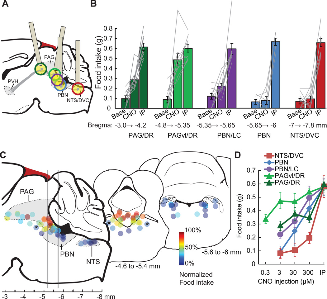
Figure 5. Cell type-selective synaptic silencing localizes a feeding circuit.
A) Schematic of descending axon projections from PVHSIM1 neurons, which express hM4Dnrxn. These axon projections were targeted in separate animals for spatially defined synaptic silencing by intracranial microinjection of CNO. Estimated precision of microinjections (based on ±500 µm) at each site is indicated by colored circles (for clarity, cannula schematic is not shown for ‘purple’ injection site). Injection cannula was angled for PAG targeting in order to avoid the sinus confluens (blood vessel, dark red).
B) Pre-injection baseline (Base) and evoked feeding response (1 h) from regions targeted by intracranial injections (CNO, 3 µM, typically bilateral) at distinct anterior-posterior positions. The most efficacious feeding responses were observed with microinjections into the PAGvl/DR (light green), which was similar to food consumption evoked by intraperitoneal injection of CNO (IP). Intracranial microinjections that were more ventral or dorsal than the indicated areas were not included in the analysis.
C) Schematic of sagittal section showing the location of injection sites, which are color coded to reflect food intake evoked after intracranial 3 µM CNO microinjection, normalized to feeding after IP injection. Two transverse sections are displayed, at the level of PAGvl/DR and the caudal PBN, with the positions of multiple injection sites projected onto a coronal diagram. Bilateral injections are displayed as identically color coded pairs. Asterisks denote bilateral microinjection site at the pedunculopontine tegmental nucleus (PPTg), which did not evoke substantial food intake.
D) Dose response for CNO microinjections at different injection sites. Selectivity is reduced with increasing CNO dose, such that microinjection sites outside the PAGvl/DR region can evoke feeding. Values are means ± SEM.

