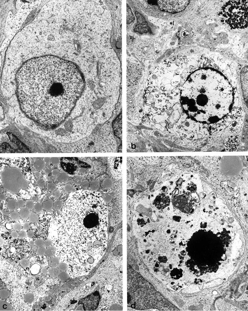
FIG. 7.
Degenerating cells in morphine-treated explants at 7 to 10 days in vitro. With progressive degeneration and loss of cellular morphology, it can be difficult to identify Purkinje cells with certainty. (a) A Purkinje cell with a deficit in cytoplasmic organelles and abnormally dense marginal heterochromatin (8,770×). (b) A dying cell surrounded by astrocytic processes containing numerous intermediate filaments (asterisks) (6,150×). (c) A degenerating cell with accumulated lipid and glycogen in the cytoplasm and partial destruction of the nuclear membrane. This cell had remnents of hypolemmal cisternae (not shown) (5,390×). (d) A large pyknotic cell positioned within the Purkinje cell layer that is difficult to identify with certainty (6,750×).

