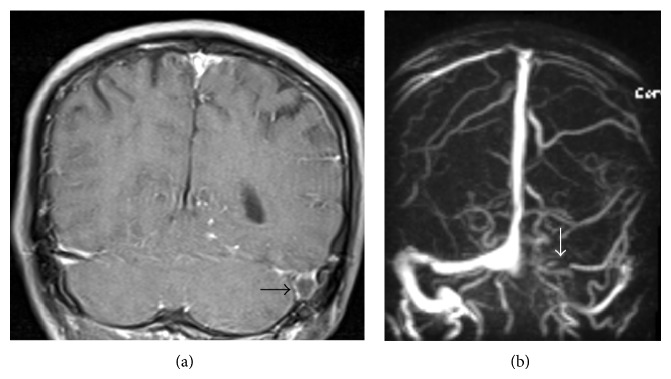
Figure 2.

Dural venous sinus thrombosis as an only imaging evidence of tuberculous meningitis in a 45-year-old male who presented with headache and cerebrospinal fluid PCR positive for Mycobacterium tuberculosis. (a) Coronal postcontrast T1-weighted MR image demonstrates a filing defect within dilated left sigmoid sinus (black arrow). (b) MR angiogram reveals nonvisualization of transverse and sigmoid sinuses in left side (white arrow).
