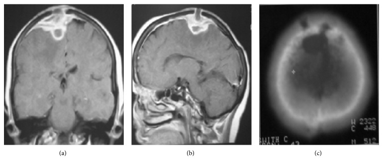
Figure 8.
Tuberculous abscess with epidural and subdural empyema and calvarial osteomyelitis. Coronal (a) and sagittal (b) postcontrast T1-weighted MR images demonstrate epidural and subdural collections over the bifrontal cerebral convexities (more on the right side) with intraparenchymal and calvarial extension. Peripheral edema and irregular marked enhancement of the lesion as well as dural enhancement are evident. The bony destructive lytic lesions are seen in the bone window CT image (c).

