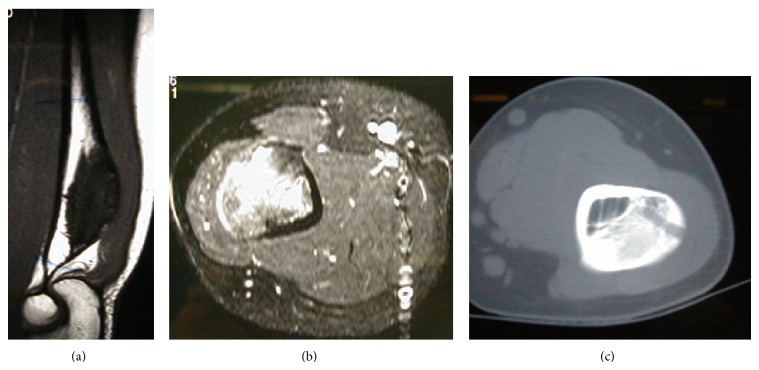
Figure 2.
The MRI images show an exostosis arising from the posterior surface of the distal humerus. This lesion measured approximately 6 cm in its long axis terminating immediately proximally to the level of the olecranon and with involvement of the underlying medulla. You can see the sign of the biopsy (c).

