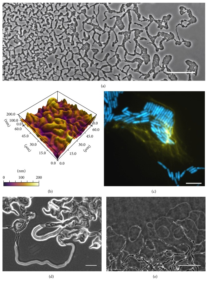
Figure 1.
Stigmergic self-organisation of bacterial communities. (a) Pseudomonas aeruginosa interstitial biofilm imaged using phase contrast microscopy depicting the emergent pattern formation. At the advancing edge are rafts of cells that initiate biofilm expansion, behind which there is an interconnected lattice-like network of cellular trails. Scale bar indicates 50 μm. (b) 3D rendered image of the interconnected furrow network underlying the P. aeruginosa interstitial biofilms imaged using atomic force microscopy (AFM) within the lattice-like network. Height scale is relative. (c) P. aeruginosa expressing cyan fluorescent protein (CFP; blue) interstitial biofilms were grown on media supplemented with the cell impermeant nucleic acid dye TOTO-1 to visualize eDNA (yellow) and imaged using OMX BLAZE wide-field microscopy. Scale bar indicates 5 μm. Swarming communities of (d) Pr. vulgaris and (e) M. xanthus grown on semisolid nutrient media and imaged using phase contrast microscopy revealing the phase bright trails routinely observed at the leading edge. Scale bar is 100 μm.

