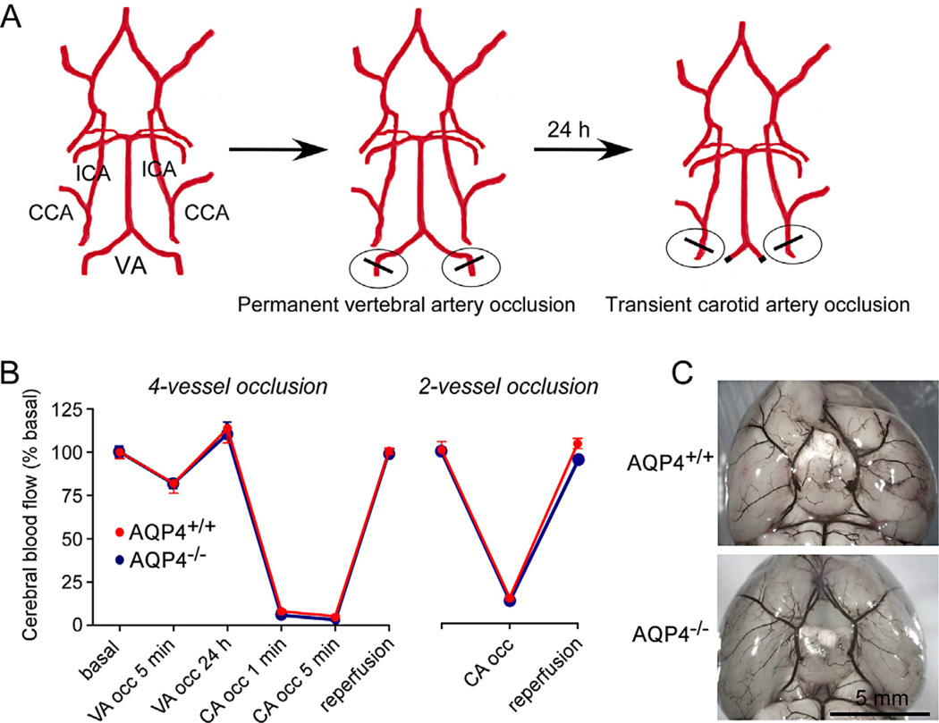
Fig. 1.
4-Vessel occlusion model of severe global cerebral ischemia in mice. (A) Diagram of approach showing permanent vertebral artery (VA) occlusion produced by cautery at 24 h before transient bilateral common carotid artery (CCA) occlusion. ICA: internal carotid artery. (B) Cerebral blood flow measured by Doppler at indicated times in 4-vessel (left) and 2-vessel (right) models (mean ± S.E., 4 mice per group). (C) Cerebrovascular anatomy visualized following intravenous ink injection.

