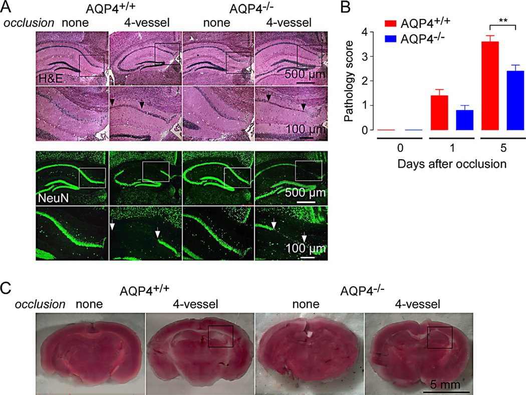
Fig. 3.
Reduced neuronal injury in AQP4-deficient mice following 4-vessel occlusion. (A) Hematoxylin and eosin-stained sections through hippocampus in control mice and at 5 days after 4-vesssel occlusion (top). Higher magnification of boxed areas shown. NeuN (neuronal marker) immunofluorescence (bottom). Micrographs representative of 5 mice studied per condition. (B) Pathological score (see Methods, score 0 without pathology) deduced from analysis of hippocampal sections (S.E., 5 mice per group, **P < 0.05). (C) TTC staining of freshly cut brain of control mice and at 5 days after 4-vessel occlusion. Hippocampus region indicated by box. Representative of TTC staining from 5 mice.

