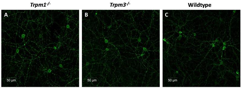
Figure 4.
Melanopsin expression in Trpm1−/− and Trpm3−/− mice. Confocal images, showing the levels of melanopsin immunoreactivity observed in Trpm1−/− (A), Trpm3−/− (B) and normal wildtype whole mounted retina (C). Note that no differences were observed, with levels of expression, number of cells and distribution of melanopsin expressing cells similar for all strains of mice.

