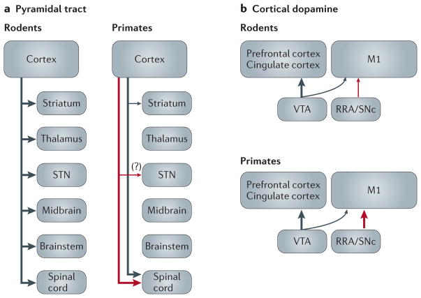
Figure 1. Highly simplified diagram of key anatomical differences between rodents and primates with regard to pyramidal tract neurons (a) and the sources of dopamine innervation of prefrontal/cingulate and M1 cortices (b).
Red and black arrows are used to show projections from distinct neuronal populations. The thickness of arrows in these diagrams indicates relative differences in the extent of target innervation by the afferent inputs. For simplicity, some corticostriatal neuron subtypes are not shown. M1, primary motor cortex; RRA, retrorubral area; SNc, substantia nigra pars compacta; STN, subthalamic nucleus; VTA, ventral tegmental area.

