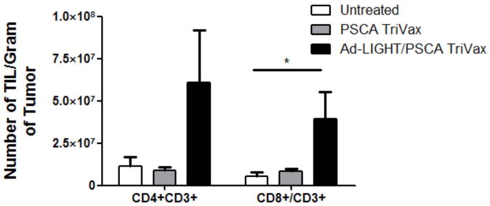
Fig. 2.

Increase in intratumoral CD4+ and CD8+ T cells following forced expression of membrane bound LIGHT in a prostate cancer tumor model. (A) Tumor infiltrating lymphocytes were released from untreated or treated tumors 7 days after Ad-Control or Ad-LIGHT injection. Cells were stained with CD4, CD8 and CD3 Ab and analyzed via flow cytometry. The number of TIL/gram of tumor from CD8+/CD3+ and CD4+/CD3+ T cells were significantly higher in Ad-LIGHT treated mice compared to untreated. (p<0.05, one-way ANOVA). (B) The number of CD4+CD25+Foxp3+ Tregs per gram of tumor were not significantly differently, despite the increase in total number of infiltrating lymphocytes in the Ad-LIGHT samples. Shown is the average number of FoxP3+ TIL (±SD) from 5 treated mice/group. Data are representative of two individual experiments.
