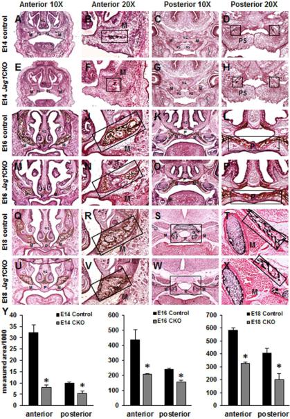
Figure 3. Diminished ossification in Jag1CKO maxillary and palatine bones.
Von Kossa staining was performed on anterior and posterior frozen sections through the developing maxilla at E14 (A-H), E16 (I-P), and E18 (Q-X) in control and Jag1CKO embryos. Reduced areas of ossification in both the lateral maxillary bone (M) and medial palatine bone (P) were revealed in Jag1CKO embryos when compared to controls (boxes and graphs). Columns, mean area obtained from 3 separate experiments; bars, SEM; *=p<0.05. M=maxilla, PS=palate shelf, P=palate.

