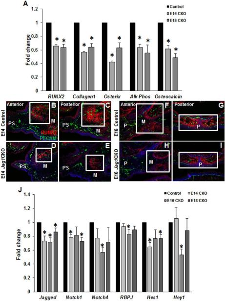
Figure 4. Aberrant osteoblast development and differentiation in Jag1CKO maxillas.
qPCR revealed a decrease in the induction of osteoblast differentiation genes at both E16 and E18 in Jag1CKO whole maxillary mRNA when compared to control littermates at the same developmental time point (A). Columns, median fold change obtained from 3 separate experiments; bars, SEM; *=p<0.05. Immunohistochemistry (B-I): Frozen sections from the anterior and posterior developing maxilla in control and Jag1CKO were stained with an early marker of osteoblast differentiation, RUNX2, and endothelial cell marker, PECAM. The density of RUNX2 staining (boxes) was reduced in Jag1CKO maxillas at E14 (D, E) and E16 (H, I) when compared to control littermates (B, C, F, G). n=3 for each time point. M=maxilla, PS=palate shelf, P=palate. qPCR was used to compare the expression of Jagged1 ligand, Notch 1 and 4 receptors, and downstream effectors: RBPJ, Hes1, and Hey1 in Jag1CKO whole maxillary mRNA at E14, E16, and E18 to control littermate whole maxillary mRNA at the same developmental time point (J). Columns, mean fold change obtained from 3 separate experiments; bars, SEM; *=p<0.05.

