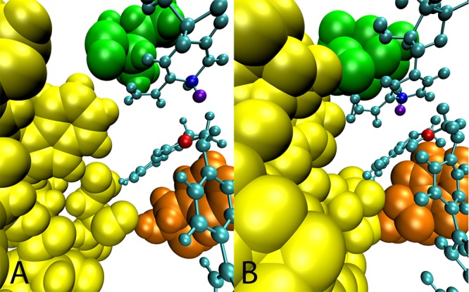
Figure 5.

Structural differences between the ecDHFR and hsDHFR active sites. View of the equilibrated structure of the (A) ecDHFR and (B) hsDHFR system. The ligands are cyan, the hydrides are violet, the hydride donors are blue, and the hydride acceptors are red. Residues of the Met20 loop are yellow, I14/17 is green, and residue F31/35 is orange.
