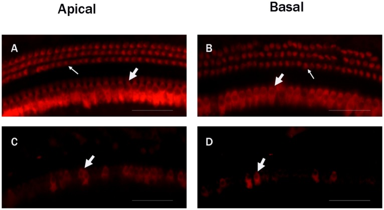
Fig 1. Fluorescent immunohistochemistry images of myosin VII7a-labeled hair cells in the cochleae of the C57BL/6J mice.

A: The apical turn in the control group. B: The basal turn in the control group. Three rows of outer hair cells (Thin white arrow) and one row of inner hair cells (Thick white arrow) in the control group were complete and arranged orderly. C: The apical turn in the experimental group at the first day following drug administration. D: The basal turn in the experimental group at the first day. Most of the outer hair cells and part of the inner hair cells in the experiment groups were destroyed. Scale bar = -25μm.
