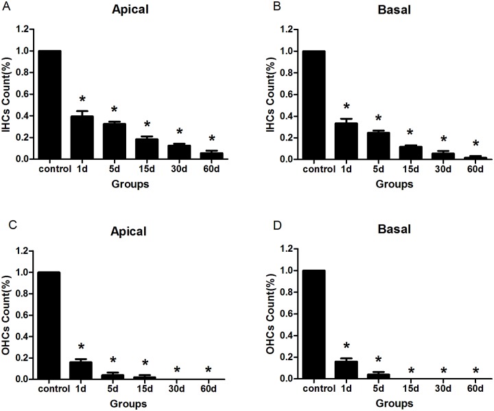
Fig 2. The changes of hair cells in the cochleae of the animals treated with kanamycin and furosemide.
A: Inner hair cells counts from the apical turn. B: Inner hair cells counts from the basal turns. C: Outer hair cells counts from the apical turns. D: Outer hair cells counts from the basal turns. Four animals were included in each group. In each cochlea two representative locations were counted. It showed that the drug administration successfully destroyed almost all hair cells in the cochlea. Asterisks indicated significant (P <0.05) difference between the experimental and the control groups.

