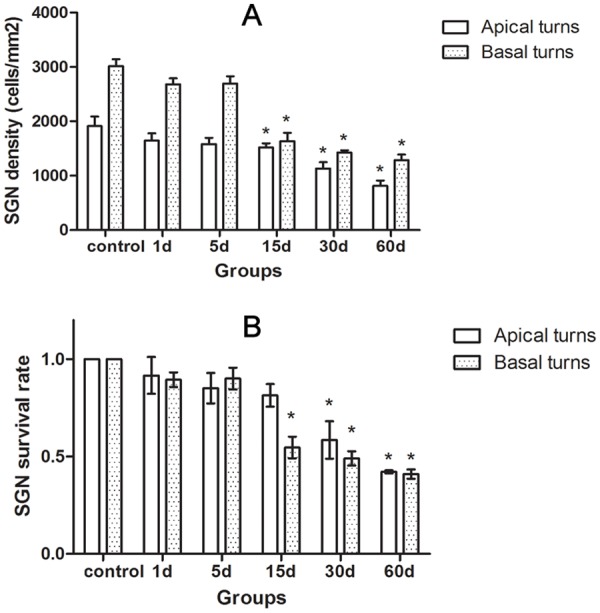
Fig 4. A: The changes of SGC density in the cochleae of the animals treated with kanamycin and furosemide.

B: Changes of SGC survival rate in the cochleae of the animals treated with kanamycin and furosemide at survival time. It showed that the SGC degeneration in the cochlea following hair cell loss was progressive. Asterisks indicated significant (P < 0.05) difference existed compared to the control group.
