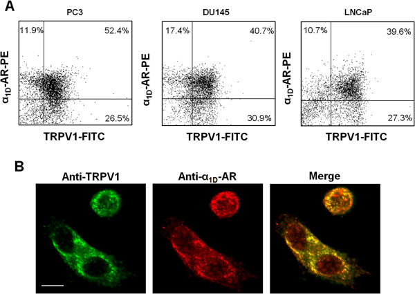
Figure 1.

α 1D -AR and TRPV1 are co-expressed and co-localized in prostate cancer cells. (A) FACS analysis was performed in PC3, DU145 and LNCaP cells double-stained with anti-TRPV1 and anti-α1D-AR Abs followed by respective secondary Abs. (B) Confocal microscopy analysis was performed using PC3 cells grown for 24 h on poly-L-lysine coated slides, permeabilized and double-stained with anti-TRPV1 and anti-α1D-AR Abs, followed by Alexa Fluor 488- and Alexa Fluor 594-conjugated secondary Abs, respectively. Bar: 20 μm. Data shown are representative of one out of three separate experiments.
