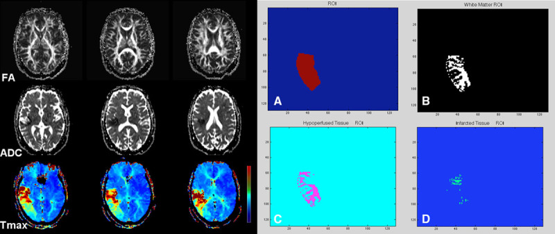
Figure 1.

A 71-year-old woman with sudden onset left-sided weakness, baseline National Institutes of Health Stroke Scale, 17. Concurrent MRA showed left M1 occlusion (not shown). Sequential aligned diffusion tensor imaging-fractional anisotropy (FA), apparent diffusion coefficient (ADC), and dynamic contrast susceptibility (DSC)-Tmax are shown. There is acute infarction in the right corona radiata and subinsular region with a large hypoperfusion deficit along the right middle cerebral artery territory on DSC-Tmax images. Aligned and coregistered maps were transferred to a Matlab program. Using DSC-Tmax, a regions of interest (ROI) was drawn over the hypoperfused region (A). After subtraction of a gray matter mask, an ROI subsuming the white matter voxels was generated (B). The FA and Tmax values were then calculated in the region of hypoperfusion (C) and infarction (D) after inclusion of an ADC map with threshold of greater and <600×10−6 mm2/s, respectively.
