
Fig. 6.
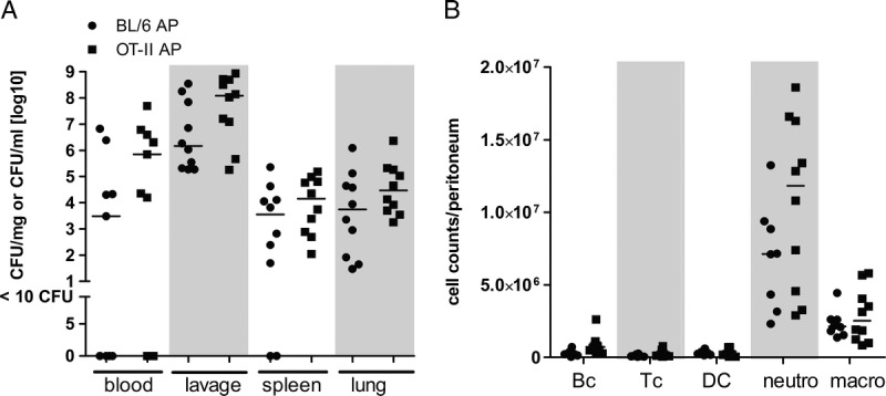
Influence of TCR signaling on bacterial clearance and peritoneal immune cell composition. OTII and C57BL/6 control mice were subjected to AP. A, After 24 h, peritoneal lavage was performed, blood was collected, and spleen and lung were recovered. Lavage, anticoagulated blood, spleen, and lung tissue homogenates were incubated on Columbia blood agar for 22 h at 37°C to determine bacterial load. Colony-forming units (CFU) were related to fluid volume or organ weight. B, Cell counts of B cells (Bc), CD4+ T cells (Tc), dendritic cells (DC), neutrophils (neutro), and macrophages (macro) were determined by flow cytometry as described in Materials and Methods. Medians are indicated. Sample sizes were nine to 10 per group. Septic OTII and WT mice were compared using the Mann-Whitney U test, showing no significant differences.