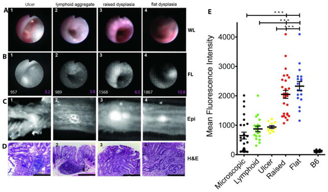
Figure 3. Focal increases of cathepsin activity emission designate dysplastic lesions in IL10−/− mice with active colitis.

(A) White light endoscopic images of lesions that correspond to (B) representative areas of focally increased NIRF emission of cathepsin activity mouse colon MFI of the lesions are shown in white fonts and SNR in magenta. In (C) is shown reflectance fluorescence image of the lesions extracted from whole mounts colon examined in A and B (660nm). (D) H&E images (x400) of the representative lesions shown in (A) & (B). (E) Cumulative plot of MFI of the lesions observed in the 15 mice examined.
