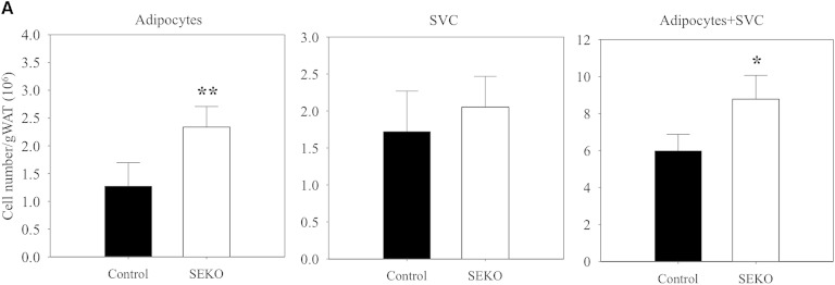
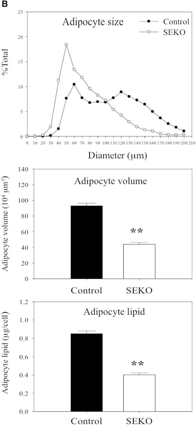
Fig. 3.
Adipocyte number and size at 8 weeks. Mature adipocytes and SVCs were isolated from visceral adipose tissue of SEKO or control mice as described in the Methods. A: Adipocyte and SVC number was estimated as described in the Methods. B: Mature adipocyte size and lipid content were analyzed using CCAP software as described in the Methods. Results are the mean ± SD of five mice. *P < 0.05; **P < 0.01 for the difference between control and SEKO mice.


