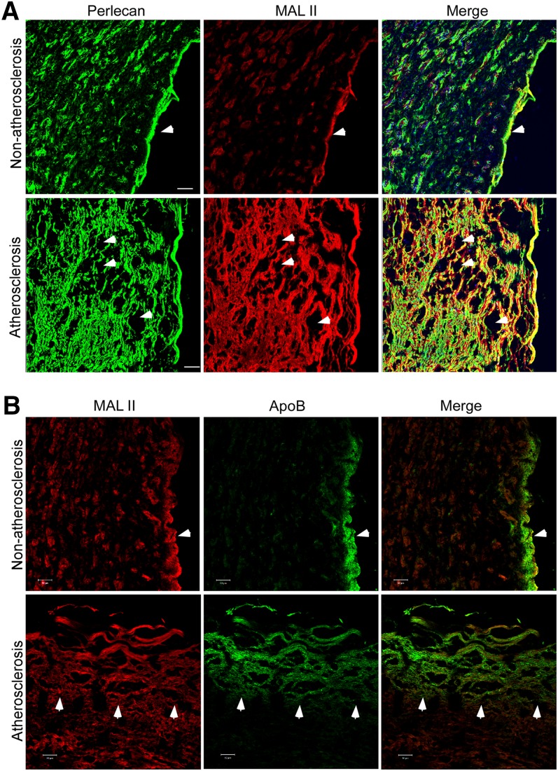
Fig. 6.
Perlecan and (α2-3)-linked sialic acid modification are overexpressed in human atherosclerotic arteries. Human normal and atherosclerotic arterial sections were stained with the perlecan-specific antibody (green) and MAL II lectin (red) (A) or the ApoB-specific monoclonal antibody (green) and MAL II lectin (red) (B), as indicated, and appropriate secondary antibodies. The images were obtained from a confocal microscope. The basement membrane in the normal tissue section (A and B, top), lipid accumulation area in the atherosclerotic section (A, bottom), and the edge of ApoB accumulation area (B, bottom) are indicated with arrows. Scale bar, 50 μm.

