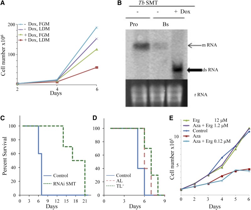
Fig. 4.
Growth of BSFs cultured in vitro with and without inhibitor treatment in FGM or LDM and Kaplan-Meier survival analysis of mice infected with and without DOX-induced TbSMT RNAi cells and with inhibitors of TbSMT. A: TbSMT RNAi strain cultured with and without DOX in FGM or LDM. B: Northern blot of TbSMT RNAi cell line. Pro, PCF; Bs, BSF. C: Survival data for T. brucei-infected mice. Solid blue line, control group (infected, wild-type BSF); dashed green line (infected, TbSMT RNAi BSF cell line). D: Survival data for T. brucei-infected mice. Solid blue line, control group (infected, wild-type BSF); red dashed line, AZA; and green-dotted line, 25-thialanosterol sulfonium salt (TL+), treated mice at 5 mg/kg. E: Rescue experiment of BSF-TbSMT RNAi cells in FGM supplemented with AZA at 1 μM (ED50 concentration) with increasing concentrations of ergosterol (Erg) as shown. For all panels, the average of three replicates is shown. Error bars are not shown because, in most cases, they approximate the data symbols.

