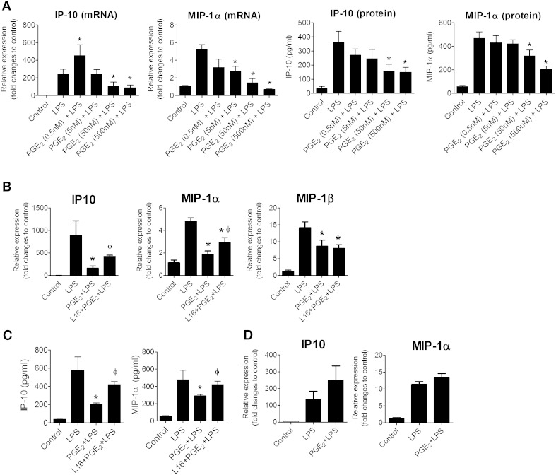
Fig. 1.
PGE2-mediated suppression of chemokines in mouse adipose tissue depends on EP4 receptor. A: PGE2 reduces IP-10 and MIP-1α expression in LPS-activated adipose tissue explants. RNA was isolated from adipose tissue explants pretreated with different concentrations of PGE2 (0.5 to 500 nM for 1.5 h) before exposure to LPS (5 ng/ml) for 9 h. The mRNA expression of IP-10 and MIP-1α for different treatment groups was expressed in relative amount produced in control. IP-10 and MIP-1α protein production in the medium was measured by ELISA and expressed in absolute values. N = 6; *, P < 0.05 versus LPS. B: Adipose tissue explants of EP4+/+ mice were treated with PGE2 (50 nM) in the presence or absence of a selective EP4 antagonist (L161,982). After 1.5 h of treatment, the cells were stimulated with LPS for 9 h. The mRNA expression of IP-10, MIP-1α, and MIP-1β in different samples was quantified. Data are expressed in relative amount produced in control. N = 6; *, P < 0.05 versus LPS; ɸ, P < 0.05 versus PGE2+LPS. C: IP-10 and MIP-1β production in the medium released from adipose tissue of EP4+/+ mice, as measured by ELISA. N = 6; *, P < 0.05 versus LPS; ɸ, P < 0.05 versus PGE2+LPS. D: PGE2 did not suppress LPS-stimulated IP-10 and MIP-1α in EP4-deficient adipose tissue. RNA was isolated from eWAT derived from EP4−/− cultured under various conditions, controls, LPS (5 ng/ml for 9 h), PGE2 (50 nM, pretreated for 2 h) + LPS (5 ng/ml for 9 h), and amounts of IP-10 and MIP-1α in different treatment groups were quantified and expressed against that of controls. N = 6. All data are expressed as means ± SEM.

