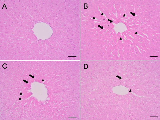
Figure 1.
Representative photographs of liver sections treated with (A) vehicle, (B) cisplatin (7.5 mg/kg), (C) cisplatin & PYC 10 (10 mg/kg) and (D) cisplatin & PYC 20 (20 mg/kg). Liver from cisplatin-treated rats showing moderate degeneration/necrosis of hepatocytes around the central vein region (open arrow head), vacuolation (closed arrow), and sinusoidal dilation (closed arrow head). H&E stain. Bar=50 µm (×400).

