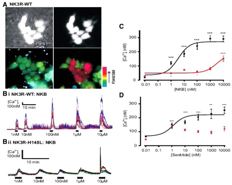
FIG. 2.
NK3R-H148L mutated receptor results in loss of function. A, Brightfield (top left) and GFP-fluorescent cells only (top right) identifying TACR3 transfected cells. Bottom left and right images depict pseudocolored representations of the 340/380 nm ratio before and after application of 10 μm NKB, respectively. B, Representative [Ca2+]i traces of WT (i) and H148L TACR3 (ii) transfected cells in response to increasing concentrations of NKB. C, NKB dose response of peak [Ca2+]i for WT (black, n = 41–53) and H148L TACR3 (red, n = 71–76). The resulting logistic fit parameters for WT were A1 = 36 ± 17, A2 = 271 ± 11, and EC50 = 3 ± 0.1 nm. NKB only induced a significant increase in [Ca2+]i at 10 μm in the H48L-mutant receptor. D, Senktide dose response of peak [Ca2+]i for WT-TACR3 (black, n = 50-57). The resulting logistic fit parameters were: A1 = 68 ± 20, A2 = 221 ± 11, and EC50 = 1 ± 1 nm. Responses to senktide on the H148L-mutant TACR3 (red, n = 50–90) were not statistically significant from nontransfected cells. A minimum of five experiments were performed for each construct and agonist. **, P < 0.01; ***, P < 0.001, compared with nontransfected cells. Error bars represent sem values.

