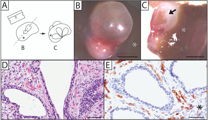
Figure 2.
Human prostate xenografts display proper growth and vascularization. (A) A drawing depicting the orientation of images shown in B and C. Human fetal prostate 200 days post-implantation into the renal subcapsular space of an immune-deficient rat host (B) was bisected (C), showing the gross appearance of ducts and stroma. Microscopically, a 7-day prostate xenograft shows normal epithelium and stromal growth (D), as well as vascularization that stain with the human CD31 endothelial marker (E). Arrow depicts ductal tissue that has canalized. Asterisk (*) indicates the location of the kidney in relation to the xenograft. Gestational age of human fetal prostate before implantation is 17 weeks (B and C), and 23.7 weeks (D and E). (Hematoxylin and Eosin staining; B, C, scale bar = 3mm; D, E, scale bar = 100 μm).

