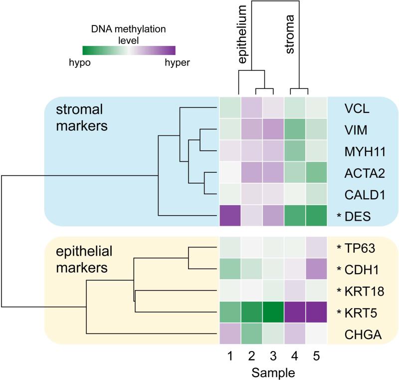
Figure 6.
DNA methylation of human prostate xenografts is different in the epithelial and stromal compartments. Heat-map of genome-wide DNA methylation analysis at promoter regions of 11 prostate marker genes specific for the epithelial (samples 1, 2, 3) and stromal (samples 4, 5) compartments. Purple color represents elevated promoter region DNA methylation, while the green color represents lowered promoter region DNA methylation relative to the mean for that promoter region. Hierarchical clustering analysis revealed that samples clustered based upon the compartment in which they are normally expressed. Sample identification is based upon age of xenograft and age of the tissue: 1: 30 days, 20 weeks gestation; 2: 30 days, 23.7 weeks gestation; 3: 90 days, 23.7 weeks gestation; 4: 30 days, 23.7 weeks gestation; 5: 90 days, 23.7 weeks gestation. Asterisk (*), promoter regions that are significantly differentially methylated by limma with Benjamini-Hochberg correction (DES, TP63, CDH1, KRT5, and KRT18).

