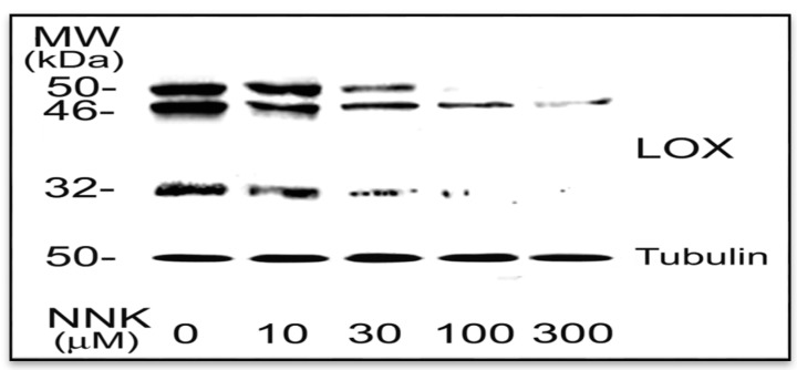
Figure 2.
NNK inhibition of LOX protein profile in treated cells. Growth-arrested RFL6 cells were treated with NNK at 0–300 µM for 48 h. Total cell proteins were extracted and aliquots of protein samples (25 µg each) were analyzed on SDS-PAGE and detected by Western blot and densitometry measurement. The 46-, 50- and 32- kDa proteins are LOX species, the bottom protein is tubulin with 50 kDa, an internal control. Experiments were repeated three times, one of which is presented here.

