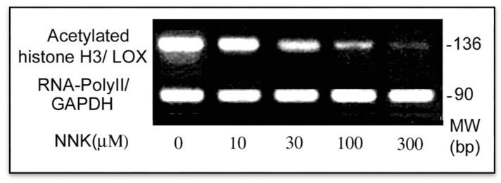
Figure 6.
Inactivation of the LOX core promoter in NNK treated cells. ChIP and PCR assays were performed to elucidate the active status of the LOX core promoter in treated cells by assessing acetylated histone H3 binding to the core promoter region. DNAs were isolated from control and NNK treated cells each with 2 × 10⁶, sonicated and immunoprecipitated with an antibody against acetylated histone H3 or RNA-PolyII. Using immunoprecipitated DNA as a template, the PCR with primer pairs as shown under Methods amplified the acetylated histone H3-bound LOX core promoter region with 136 bp, and the RNA-Poly II bound fragment of the GAPDH promoter (an internal control) with 90 bp, respectively. PCR products were analyzed on 2.2% agarose gels and densities of DNA bands measured by the 1D Scan software.

