
Figure 2.
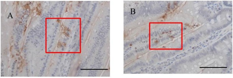
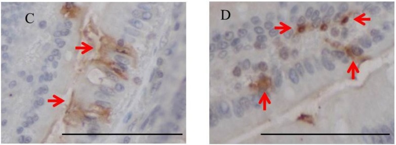
Immunohistochemistry for fucoidan in the jejunum from one-week fucoidan rat (n = 2). (A) Fucoidan stained diffusely in the epithelial cells of the jejunum (×400). Enlargement of the red frame (C). (B) Some mononuclear cells stained positively for fucoidan in the lamina propria of the jejunum (×400). Enlargement of the red frame (D). Arrows indicate positive staining for fucoidan. Bars: 50 μm.