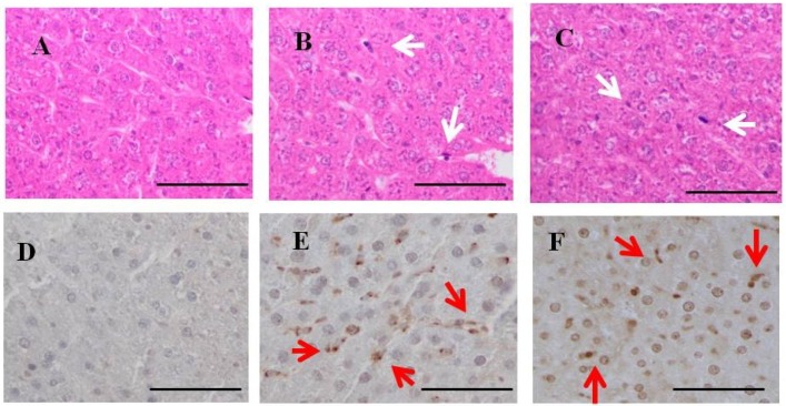
Figure 4.
H & E staining and immunohistochemistry for fucoidan in the liver of rats fed fucoidan chow or standard chow. H & E staining: (top). (A) A liver specimen from a control rat (×400); (B) A liver specimen from a one-week fucoidan rat (×400); (C) A liver specimen from a two-week fucoidan rat (×400). White arrows indicate Kupffer cells. Immunohistochemistry for fucoidan: (bottom) Fucoidan was apparent in sinusoidal non-parenchymal cells in the liver from one week-week fucoidan rat and two-week fucoidan rat, but not in control rats; (D) A liver specimen from a control rat (×400); (E) A liver specimen from a one-week fucoidan rat (×400); (F) A liver specimen from a two-week fucoidan rat (×400). Red arrows indicate positive staining for fucoidan. Bars: 50 μm.

