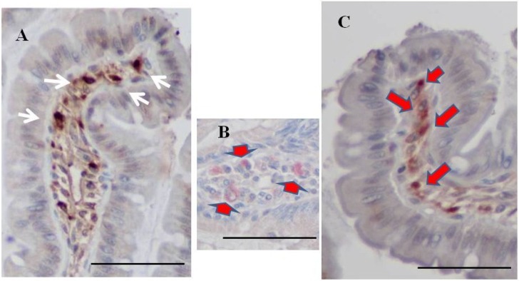
Figure 6.
Double staining for fucoidan and ED1. (A) A representative case of single staining for fucoidan. Fucoidan stained dark brown in mononuclear cells in the lamina propria of the ileum from a BBN + 2% fucoidan rat (white arrows); (B) A representative case of single staining for ED1. ED1 stained pink in round mononuclear cells in the lamina propria of the ileum from a BBN rat (arrows); (C) Some mononuclear cells in the lamina propria of the ileum from BBN + 2% fucoidan rats were positive for both fucoidan and ED1 staining. Cells were stained pink by the ED1 stain and brown by fucoidan staining (red arrows). Bars: 50 μm.

