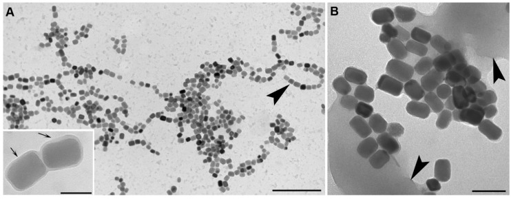
Figure 6.
Magnetosomes purified from cells of Magnetovibrio blakemorei strain MV-1. Magnetosomes purified from cells lysed using physical methods or alkaline lysis (A) with the magnetosome membrane (MM) shown in the inset (at arrows). Note that after this treatment most magnetosomes remain in chains (at arrowhead in A); Some physical-chemical methods lead to magnetosomes losing their membranes and arrangement, forming clumps due to magnetic interactions between magnetosome crystals (B). Cell debris (arrowheads in B) is generally always present in poorly washed suspensions of magnetosomes reducing purity of the preparation and potentially interfering with specific applications of the isolated magnetosomes. Scale bars = 1 μm in A (100 nm in inset), 150 nm in B.

