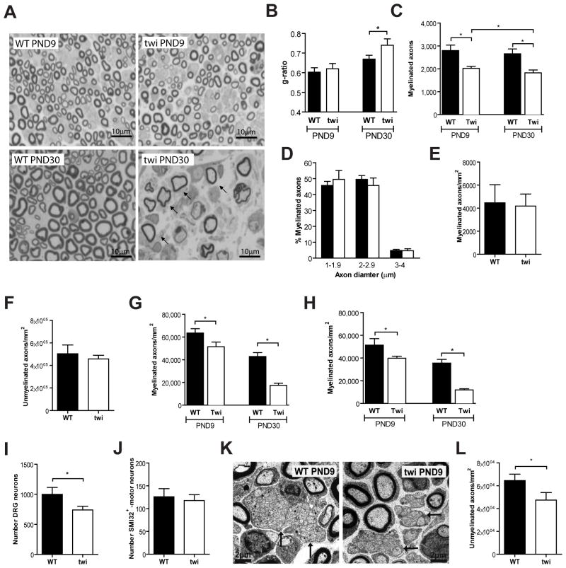
Fig. 1.
In the Twitcher sciatic nerve, a decreased axon number precedes demyelination. (A) Representative photomicrographs of transverse sections of P9 and P30 Twitcher (twi) and WT sciatic nerves are shown; arrows highlight ongoing demyelination with decreased myelin thickness. (B) g-ratio analyses in sciatic nerves of P9 and P30 WT and twi mice. (C) Total number of myelinated axons in the sciatic nerve of P9 and P30 WT and twi mice. (D) Distribution of myelinated axons according to their size in the sciatic nerve of WT and twi mice at P9. For (B–D), n=5 WT and n=5 twi mice at P9 and n=4 WT and n=6 twi mice at P30 were analyzed. (E,F) Density of myelinated (E) and unmyelinated (F) axons in sciatic nerves of P0 WT (n=5) and twi (n=3) mice. (G, H) Density of myelinated axons in the dorsal root (G) and ventral root (H) of WT and twi mice at P9 (n=4 WT and n=5 twi) and at P30 (n=4 WT and n=4 Twi mice). (I) Number of sensory neurons in WT and Twi DRG sections at P9. (J) Number of motor neurons in WT and twi mice (n=4 WT and n=6 twi mice) in spinal cord sections. (K) Representative electron microscopy photomicrographs of ultrathin sciatic nerve sections of WT and twi mice at P9; arrows highlight Remak bundles. (L) Density of unmyelinated axons in the sciatic nerve of WT and twi mice at P9 (n=5 WT and n=5 twi).

