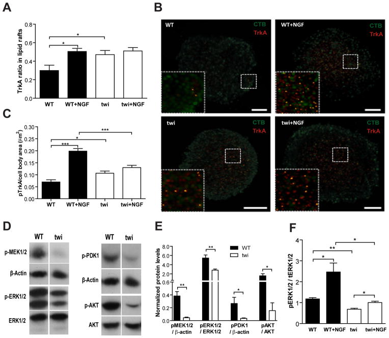
Fig. 5.
Impaired TrkA recruitment and signal transduction in Twitcher DRG neurons. (A) Ratio of TrkA localization in lipid rafts in DRG neurons of WT and Twitcher (twi) mice after 5 DIV either with (WT+NGF and twi+NGF) or without (WT and twi) NGF stimulation. (B) Representative images of the co-localization of TrkA with lipid rafts; TrkA- red, lipid rafts labeled with CTB- green in WT, WT+NGF, twi and twi+NGF DRG neurons. (C) Ratio of phosphorylated TrkA in cell bodies of DRG neurons from WT and twi mice with or without NGF stimulation. (D) Western blot analysis and (E) quantification of the MEK–ERK (left panels) and PDK1–AKT (right panels) pathways in WT and twi DRG neurons. (F) Quantification of ERK activation in WT and twi DRG neurons with or without NGF stimulation.

