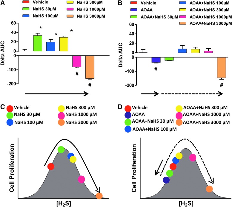
FIG. 17.
Bell-shaped effect of the fast-acting H2S donor NaHS on the proliferation of HCT116 cells in the absence or presence of AOAA. (A, B) show the effect of various concentrations of NaHS on proliferation. The HCT116 cell proliferation was assessed in real time for approximately 72 h using the xCELLigence system as described (117), and the effect of the H2S donor was calculated as the change in the area under the curve (0–72 h) relative to the proliferation of vehicle-treated cells. In this analysis, the ΔAUC of the cells in the presence of vehicle only is defined as zero. Columns that are oriented in the positive direction represent increases in proliferation in response to the H2S donor compared with vehicle (*p<0.05); negative columns represent the inhibitory effect of NaHS on cell proliferation (#p<0.05). (C, D) show the interpretation of the findings based on a bell-shaped dose-response model. (A, C) as well as (B, D) are color-coordinated to represent identical experimental groups. See “H2S Donation vs. H2S Biosynthesis Inhibition in Cancer” for additional explanation of the model. Data represent mean±SEM of n=4 independent determinations. To see this illustration in color, the reader is referred to the web version of this article at www.liebertpub.com/ars

