Figure 4.

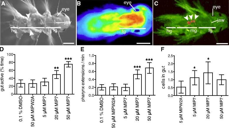
Synthetic MIP treatment increases gut peristalsis, pharynx extension and ingestion in Platynereis . (A) Differential interference contrast micrograph of 6.5 dpf Platynereis. White arrowheads indicate muscular contraction in the gut. (B) Calcium-imaging with GCaMP6 in 6.5 dpf Platynereis highlights muscular pharynx extension in the foregut. (C) Fluorescent micrograph of 7 dpf Platynereis with AF488 filter. White arrowheads indicate autofluorescent Tetraselmis cells in the gut. All images are dorsal views, with head to the right. (D) Gut activity as percentage of time in MIP-treated versus control 6.5 dpf Platynereis. (E) Number of pharynx extensions per minute in MIP-treated versus control 6.5 dpf Platynereis. (F) Number of Tetraselmis marina algae cells eaten per larvae in 30 min in MIP-treated versus control 7 dpf Platynereis. (D-F) Data are shown as mean +/- 95% confidence interval, n = 60 larvae. p-value cut-offs based on unpaired t-test: *** <0.001; ** < 0.01; * <0.05. MIPW2A is a control non-functional MIP peptide in which the two conserved tryptophan sites are substituted with alanines. Scale bars: 50 μm. Abbreviations: hg, hindgut; mg, midgut; fg, foregut.
