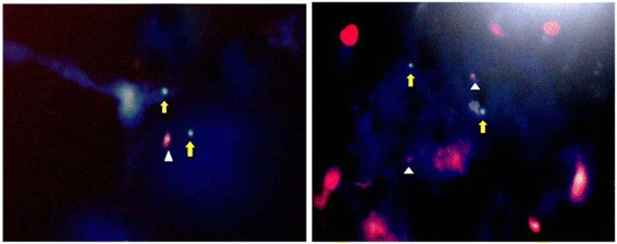
Figure 3.

FISH images of formalin fixed paraffin embedded sections of BAC. Red signals (white arrow heads) are indicative of the p53 gene probe, while green signals (yellow block arrows) are indicative of centromeric probe of chromosome 17. The blue color is that of the DAPI counter stain. The presence of a single red signal per cell reflects the presence of a single copy of the p53 gene, i.e. p53 deletion (left), right photo represents normal cell.
