Figure 7.
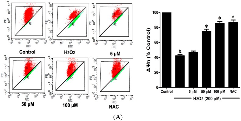
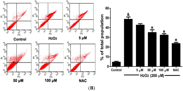
DSC inhibited H2O2-induced mitochondrial membrane potential (ΔΨm) loss and apoptosis in HUVEC. HUVEC were incubated with indicated concentrations of DSC or NAC for 4 h, then stimulated with H2O2 (200 μM) for 2 or 12 h; the ΔΨm (A) and apoptosis (B) were measured by flow cytometry, respectively. Data shown are means ± SEM, & p < 0.05 compared with unstimulated cells, * p < 0.05 compared with H2O2-stimulated cells. Data were from at least three independent experiments, each performed in duplicate.


