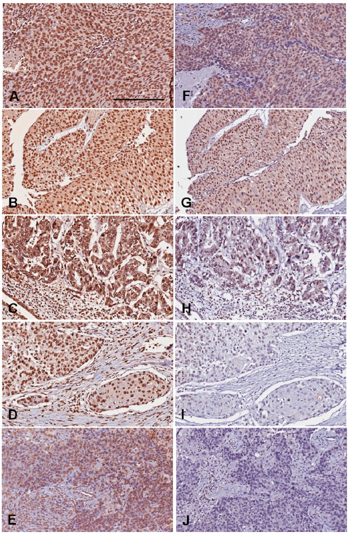
Figure 2.
Representative photography of 5-MeC and DNMT1 immunohistochemistry. The 5-MeC photography in A–E; The DNMT1 photos in F–J. (A,F) High-grade urothelial carcinoma showing high levels of both 5-MeC and DNMT1; (B,G) Low-grade urothelial carcinoma showing high levels of both 5-MeC and DNMT1; (C,H) High-grade urothelial carcinoma with glandular differentiation, showing high 5-MeC but low DNMT1 levels. (D,I) High-grade urothelial carcinoma with squamous differentiation, showing low levels of both 5-MeC and DNMT1; (E,J) High-grade urothelial carcinoma showing low levels of both 5-MeC and DNMT1. Bar = 200 µm.

