Figure 5.
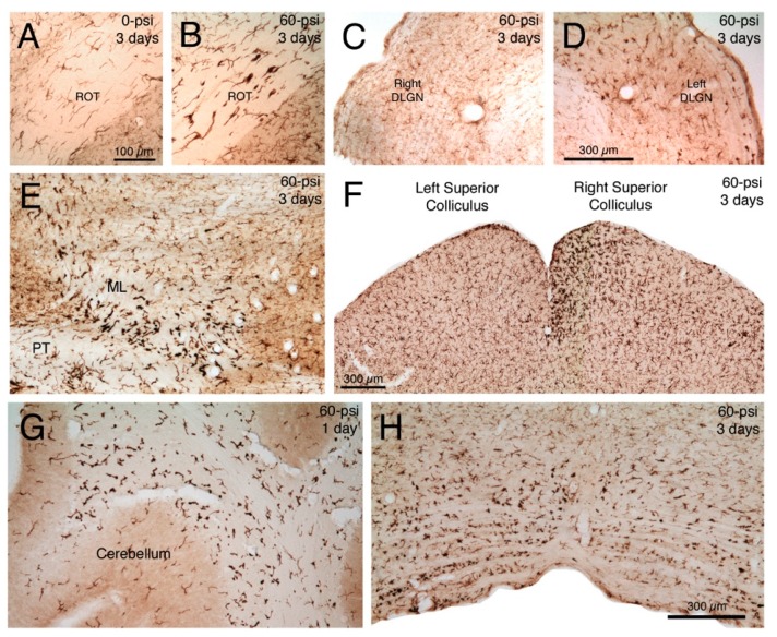
Transverse sections immunostained for ionized calcium-binding adapter molecule-1 (IBA1), showing: Normal microglia in right optic tract (ROT) three days after a left cranial 0-psi blast (A); activated microglia in the ROT three days after a left cranial 60-psi blast (B); normal microglia in left dorsal lateral geniculate nucleus (DLGN) three days after a left cranial 60-psi blast (C); activated microglia in the right DLGN three days after the same left cranial 60-psi blast (D); activated microglia in the left medial lemniscus (ML) and pyramidal tract (PT) at the level of the mesencephalon three days after left cranial 60-psi blast (E); normal microglia in left superior colliculus and activated microglia (especially medially in the upper layers) in the right superior colliculus three days after a left cranial 60-psi blast (F); activated microglia in the deep cerebellar white matter one day after left cranial 60-psi blast (G); and activated microglia in basal medulla three days after a left cranial 60-psi blast, particularly on the left side (H). Note that normal microglia (A,C) immunolabel lightly for IBA1, and have a small cell body and thin processes. By contrast activated microglia immunolabel intensely for IBA1, and have more prominent perikarya that possess thick short processes. Images A and B are at the same magnification as one another, images C and D are at the same magnification as one another, and images E, G and H are at the same magnification as one another.

