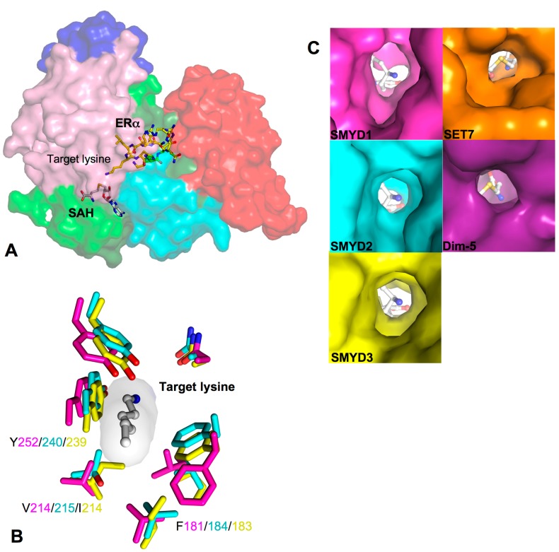
Figure 5.
Target lysine access channel. (A) Surface representation of overall SMYD2–ERα structure. ERα peptide, SAH, and target lysine are indicated; (B) Superposition of the lysine access channels. SMYD residues are represented by sticks with the carbon atoms colored according to the scheme in Figure 1C. Target lysine is colored in white; and (C) Surface representation of the lysine access channel of SMYD1–3, SET7, and Dim-5. SAH or SFG is depicted by sticks with the carbon atoms colored in white.

