Figure 5.
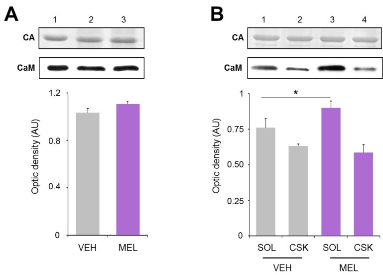
Calmodulin content in hippocampal slices treated with Melatonin. Hippocampal slices were incubated during 6 h with either the vehicle (VEH) or melatonin (MEL). Calmodulin (CaM) in the homogenates (A); and in the soluble (SOL) and cytoskeletal fractions (CSK) separated by centrifugation (B); was determined by Western blot. Upper panels show Carbonic Anhydrase (CA) used as external load control and stained with Coomassie blue. Representative fluorograms of CaM are shown immediately below. First lane of both gel and fluorogram from panel A was loaded with pure CA (5 µg) and CaM (1 µg), respectively. CaM was recognized with a specific CaM antibody and ECL. Histograms correspond to densitometric analysis of the bands shown in the upper panels. Results are the mean ± SEM of four densitometric scannings obtained from two independent experiments. Asterisk indicates significant differences (p < 0.05).

