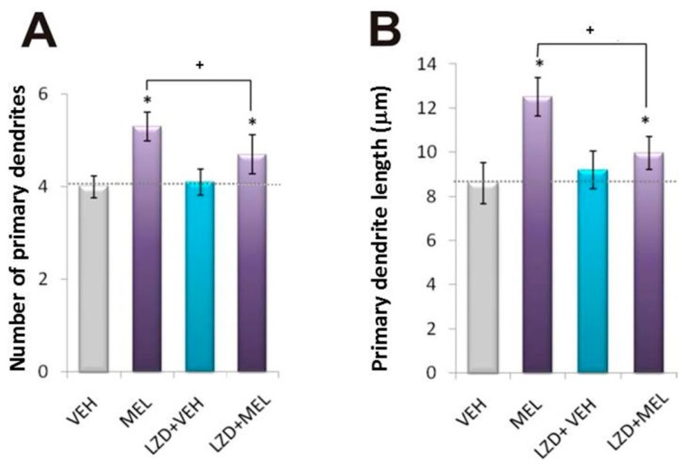
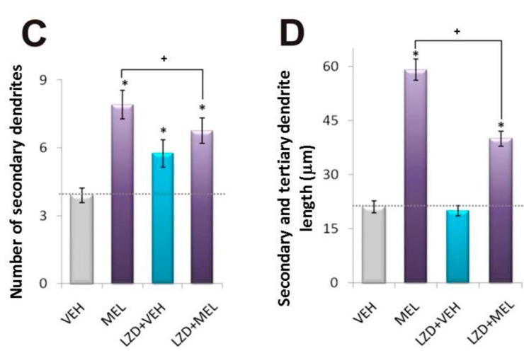
Figure 7.
Morphometric analysis of dendrite formation elicited with Melatonin in presence of Luzindole. Hippocampal slices were incubated for 6 h with either the vehicle (VEH), 100 nM melatonin (MEL), or were pre-incubated with luzindole (LZD) for 15 min followed by 6 h incubation with the vehicle (LZD + VEH) or 100 nM melatonin (LZD + MEL). After the incubation time, slices were cut into 50 µM sections and immunostained for the specific marker of dendrites MAP2. Slices were analyzed by the modified Sholl method. Results represent the mean ± SEM of one experiment of three done by quadruplicate. Asterisks show significant differences (p < 0.05). (A) Number of primary dendrites; (B) Primary dendrite length (µm); (C) Number of secondary dendrites; (D) Secondary and tertiary dendrite length (µm).


