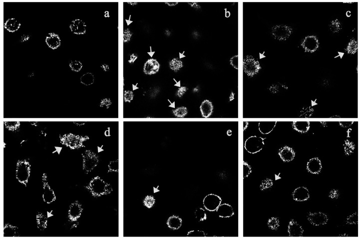
Figure 5.
Confocal fluorescence microscope observation of F-actin (labeled with rhodamin-phalloidin) in RBL-2H3 cells. The arrow represents membrane ruffling caused by F-actin rearrangement. (a) IgE-sensitized RBL-2H3 cells stimulated with PBS; (b) IgE-sensitized RBL-2H3 cells stimulated with DNP-HSA for 30 min; (c–f), IgE-sensitized RBL-2H3 cells, pretreated with 50, 200, and 800 μg/mL of TIPP or 20 μg/mL of ketotifen, then stimulated with DNP-HSA for 30 min. Magnification: 63×.

