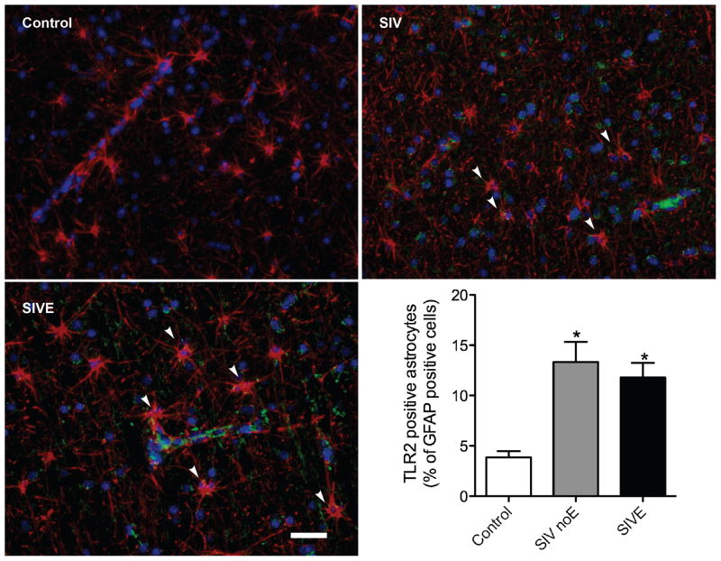
Figure 2. TLR2 expression is increased in white matter of frontal lobes of primates with SIV-induced encephalitis.
In white matter of control brain, astrocytes (GFAP, red) were generally immunonegative for TLR2 (green). The proportion of astrocytes expressing TLR2 was significantly increased in animals infected with SIV either without (SIV) or with encephalitis (SIVE). GFAP immunonegative cells, morphologically consistent with endothelial or perivascular cells, were also immunopositive for TLR2 in SIVE macaques. Scale bar = 25 μm.

