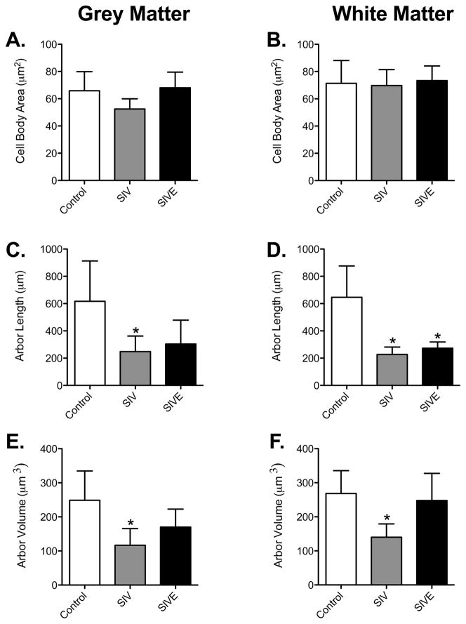
Figure 4. Decreased astrocytic arbor length and volume of animals infected with SIV.
There was no significant difference in cell body size among control, SIV noE, and SIVE animals in both grey and white matter astrocytes (A and B). In grey matter astrocytes, cell arbor was significantly decreased in SIV noE animals (C). In white matter astrocytes (D), however, cell arbors were significantly longer in astrocytes of control animals compared with astrocytes in animals infected with SIV, regardless of encephalitic status. Arbor volume was significantly decreased in astrocytes of SIV noE macaques compared with control animals. This was observed in both grey (E) and white (F) matter. Results are shown as mean ± SD (asterisk (*) indicates p <0.05 compared to controls).

