Figure 1.

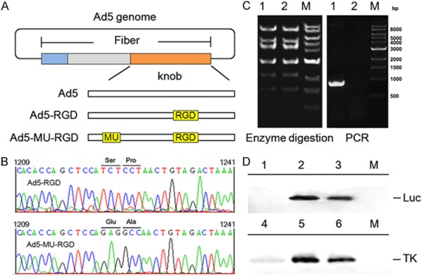
Construction and identification of fiber-mutated Ad5 vectors. A. schematic representation of Ad5 vectors carrying mutations and RGD peptide in the loops of fiber knob. B. sequencing chomatograms of the mutations (S408E, P409A) in the fiber knob. C. agarose gel electrophoresis of viral genome after restriction enzyme digestion with Hind III and PCR using specific primers. Lane 1: Ad5-MU-RGD; lane 2: Ad5-RGD; M: 1 Kb DNA ladder. D. western blot analysis of luciferase expression in 293 cells (upper) and HSV-TK expression in A549 cells (lower) after infection with Ad5 vectors. Lane 1: 293 cells; lane 2: Ad5-RGD-Luc infected 293 cells; lane 3: Ad5-MU-RGD-Luc infected 293 cells; lane 4: A549 cells; lane 5: Ad5-RGD-Luc infected A549 cells; lane 6: Ad5-MU-RGD-Luc infected A549 cells; M: pre-stained protein marker.
